Laboratory : Laboratoire de Physique des Solides
Adress : : Laboratoire de Physique des Solides, CNRS Université Paris-Saclay
1 rue Nicolas Appert, Bâtiment 510, 91405 Orsay cedex
Director of the laboratory: Pascale Foury
Supervisor : Marta de Frutos
Email adress : marta.de-frutos@universite-paris-saclay.fr
Applicant skills: A solid background in physics and/or chemistry is required together with a marked interest in experimental aspects and in particular electron microscopy, and a strong motivation for interdisciplinary physics-chemistry-biology subjects (not prerequisite knowledge).
Biominerals are materials produced by living organisms such as animal bones and teeth, mollusc shells, pathological calcifications, made from organic and inorganic compounds and organized in a multi-scale structure, from the macroscopic to nanometric level. Understanding the organization and the mechanisms of formation of these complex systems implies to analyze their chemical structure at the nanometer scale difficult to access using traditional characterization methods.
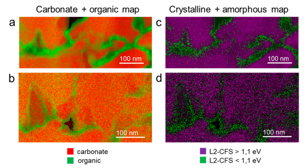
Recent studies have shown the great interest of scanning and transmission electron microscopy coupled with electron energy loss spectroscopy (STEM-EELS) for the study of these systems (1-3). This approach is based on the detection of signals induced by the interaction of the electron beam with the sample. It makes it possible to characterize the elements and chemical compounds present in the specimen through the study of the fine structure of their corresponding ionization thresholds. In addition, the use of a latest generation STEM microscope also provides information on the crystallinity of minerals (4). STEM-EELS offers the advantage of an outstanding spatial resolution (down to nanometer) compare to spectroscopic approaches based on the use of photons (X-ray absorption spectroscopies for instance). These data offer the possibility to establish chemical and structural maps of biominerals corresponding to the mineral and organic phases (2,4). For instance, STEM-EELS results on the shell of the barnacle Austromegabalanus psittacus reveals that this material is composed of irregular grains of crystalline calcite (about 200 nm in diameter) surrounded by a pellicle made of an amorphous phase rich in biomolecules (thickness about 10- 20 nm) (See Figure). These results allow to propose a model of the biomineral growth which challenges the previously established conception (4). The proposed internship aims to extend this study to other samples (current shells or fossil) in order to test the generality of the biomineralization mechanisms revealed by the study on Austromegabalanus psittacus.
The internship will take place at the Orsay Solid State Physics Laboratory within the STEM team (https://equipes2.lps.u-psud.fr/stem/) as part of a collaboration with the University of Granada (Spain).
The candidate will actively participate in different aspects of the project. He/she will be trained in STEM-EELS approaches on a latest generation microscope, the microscope Chromatem (https://equipes2.lps.u-psud.fr/stem/chromatem/). He/she will carry out measurements on biomineral samples. The acquired data will be processed by statistical analysis approaches to improve the detection limit and extract of spectral signatures of interest.
Techniques used during the internship: scanning transmission electron microscopy (STEM) coupled with electron energy loss spectroscopy (EELS) on a last generation monochromated STEM microscope.
(1) Nanoscale Analysis of Randall’s Plaques by Electron Energy Loss Spectromicroscopy: Insight in Early Biomineral Formation in Human Kidney. Gay et al, ACS Nano (2020) 14, 1823−1836; DOI: 10.1021/acsnano.9b07664
(2) Chemical and Structural Insights of the Nano Organo–Mineral Interfaces in Growing Abalone Nacre. Ajili, et al, (2023) Chemistry of Materials, 35(15): 6059–6069. DOI: https://doi.org/10.1021/acs.chemmater.3c01169
(3) Inorganic phosphate in growing calcium carbonate abalone shell suggests a shared mineral ancestral Precursor. Ajili et al, Nat. Comm. (2022) 13, 1496; DOI: 10.1038/s41467-022-29169-9
(4) Nanoscale Analysis of the Structure and Composition of Biogenic Calcite Reveals the Biomineral Growth Pattern. de Frutos et al, ACS Nano (2023) 17, 2829-2839; DOI: 10.1021/acsnano.2c11169