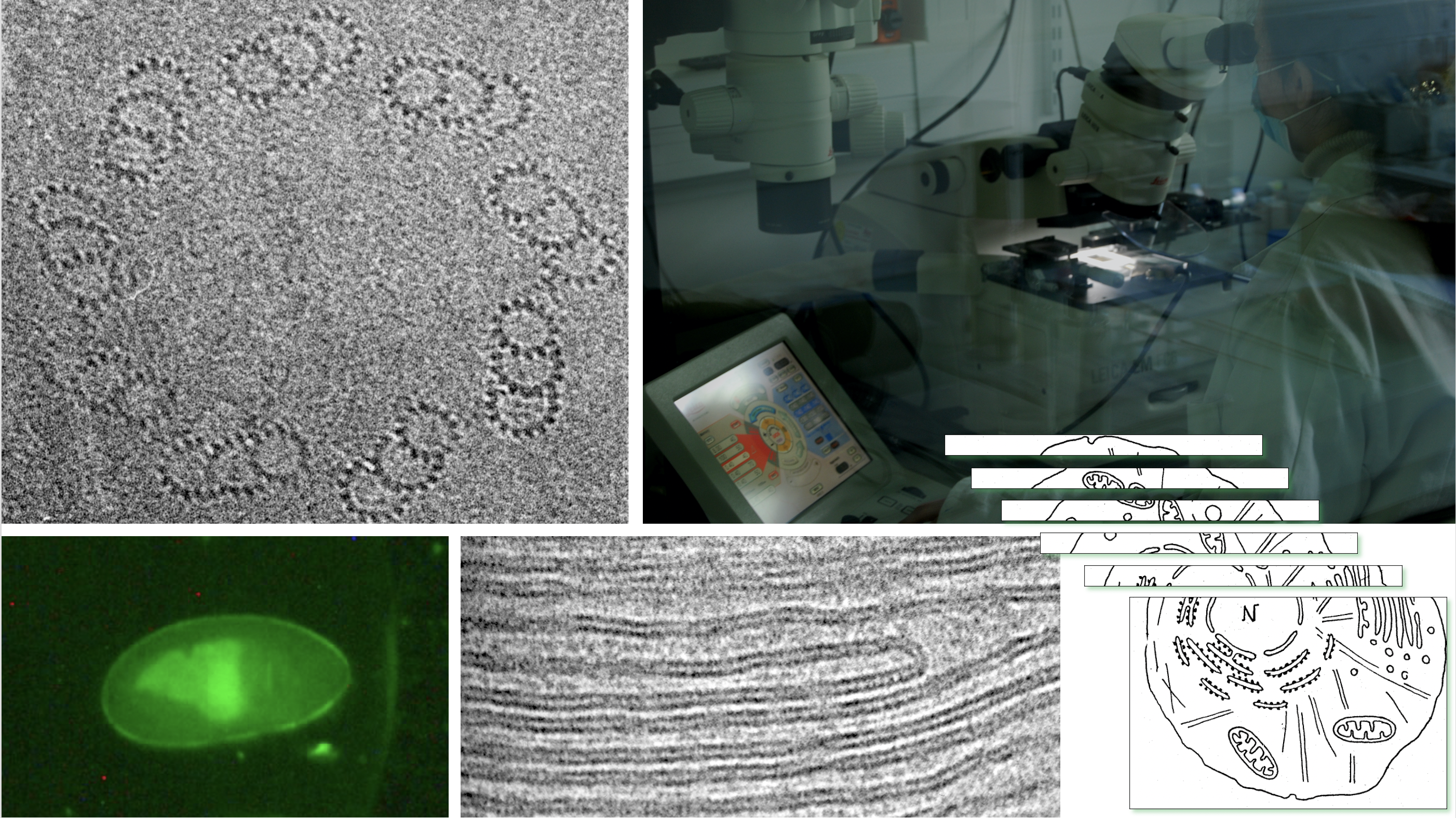
 Cryo electron microscopy of vitreous sections @ LPS
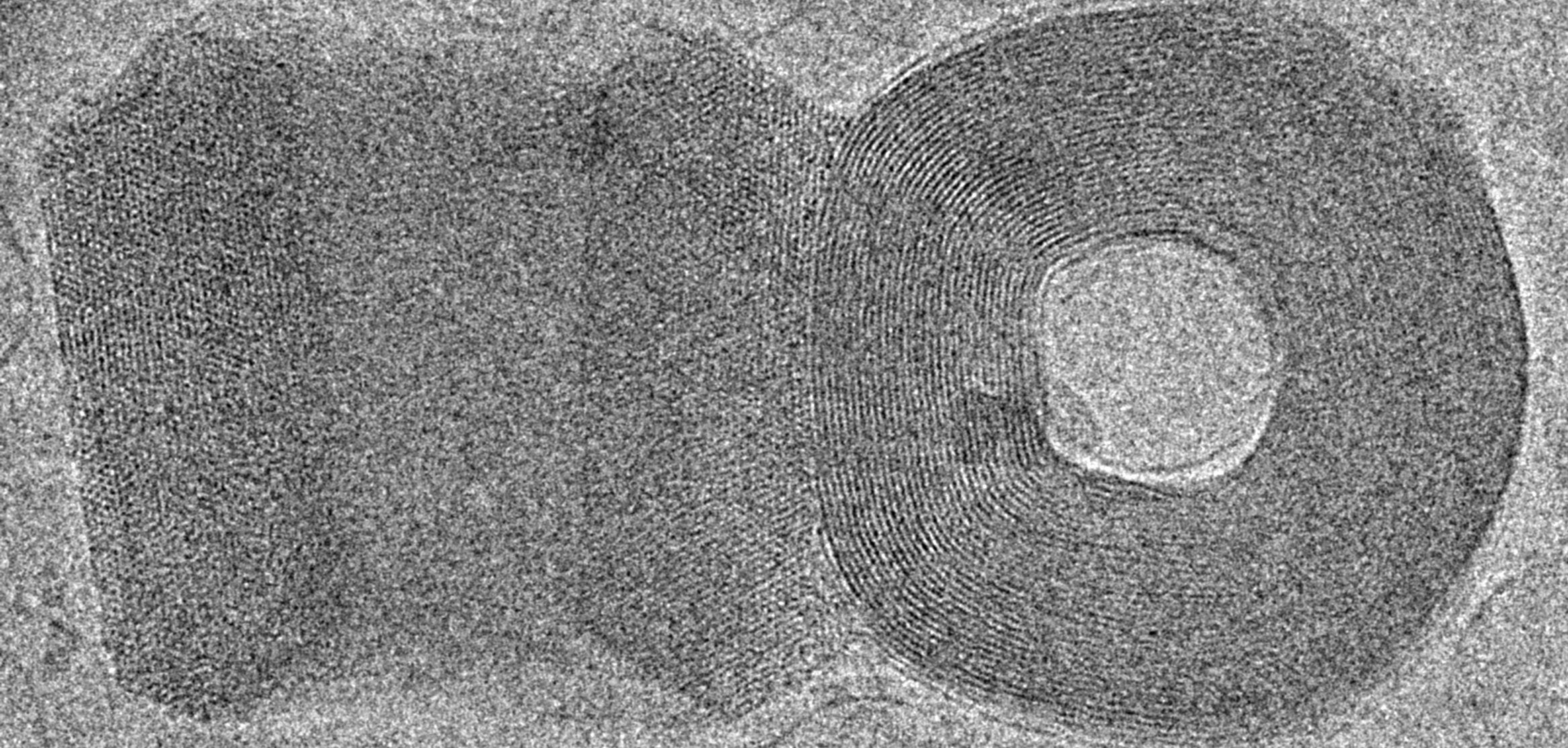 DNA toroids imaged by cryo electron microscopy. Photo Kahina Vertchik.
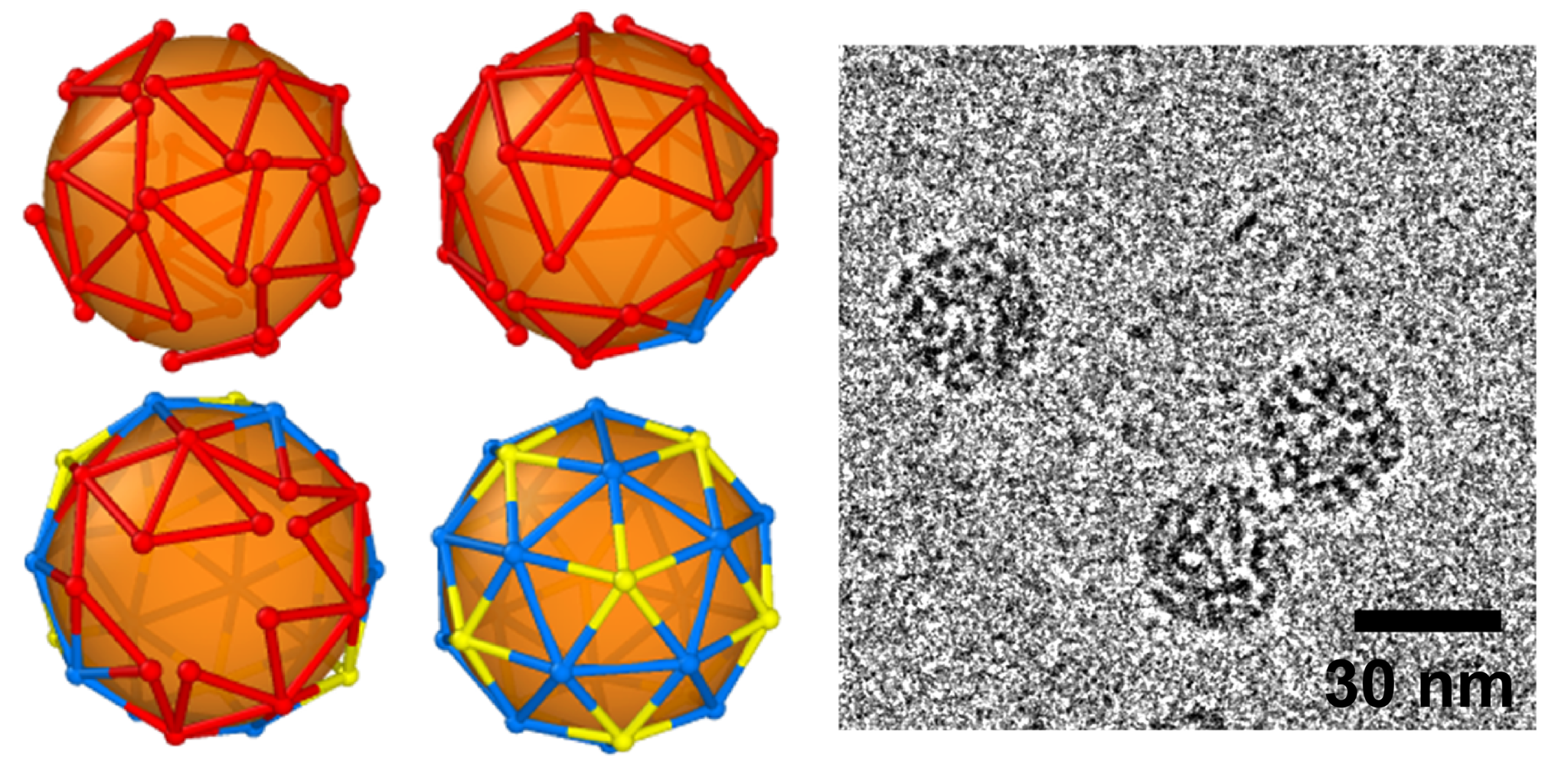 Monte Carlo simulations showing the self-organization of a viral shell (left) and cryotransmission electron micrograph of viruses assembled in vitro
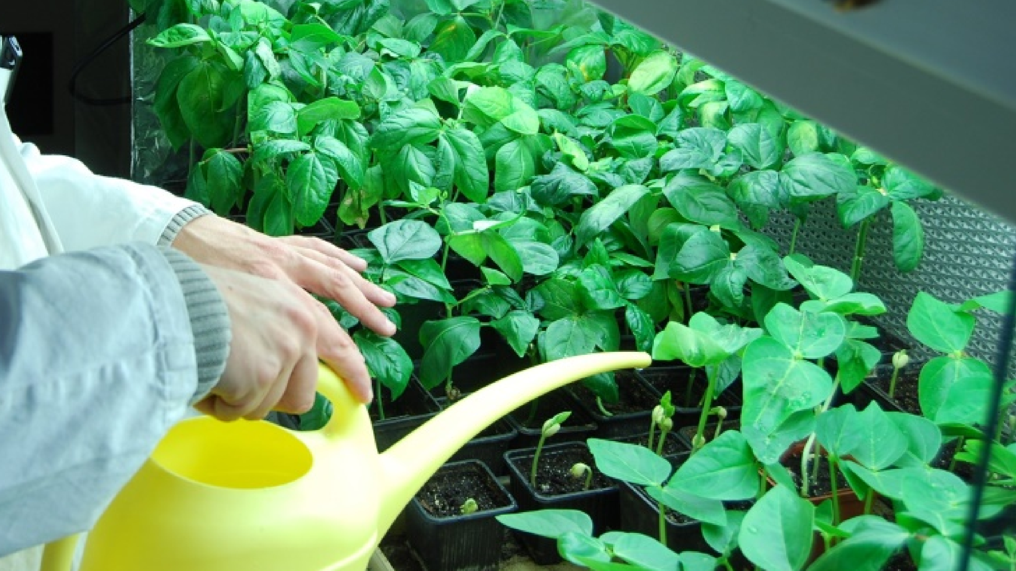 Growing cowpea for virus purification
 ESRF synchrotron
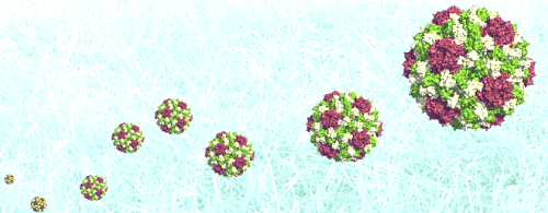 RNA packaging in viral capsid
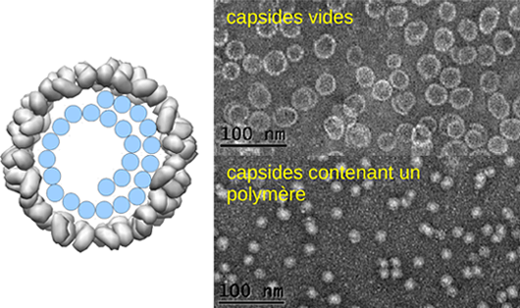 Packaging polymers in viral capsids
 Liquid crystalline nucleosomes in solution
 Cryo electron microscopy of vitreous sections @ LPS
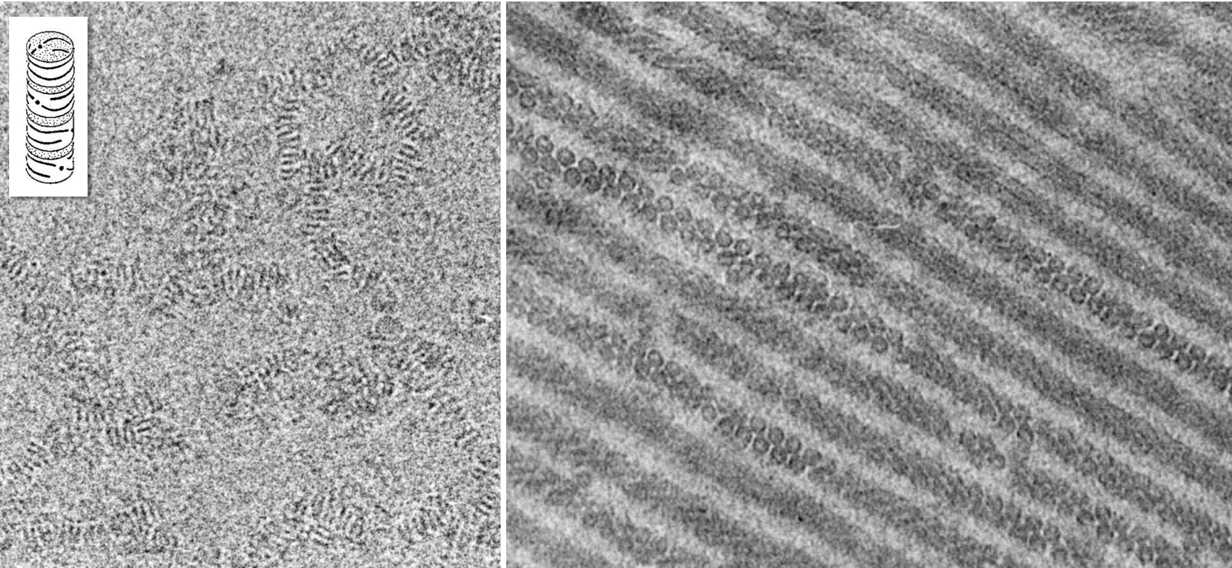 Cryo electron microscopy imaging of nucleosomes in solution
 SOBIO team
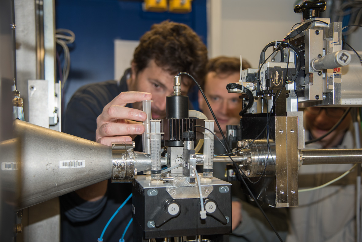 Stopped-flow coupled with synchrotron X rays
